

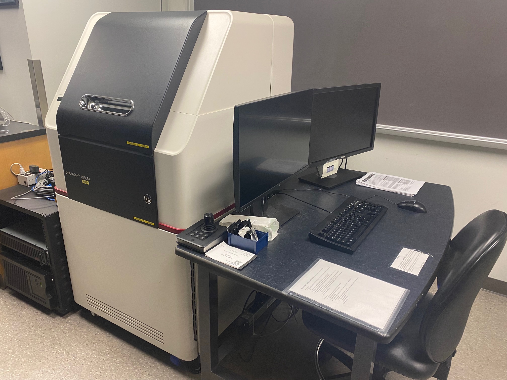
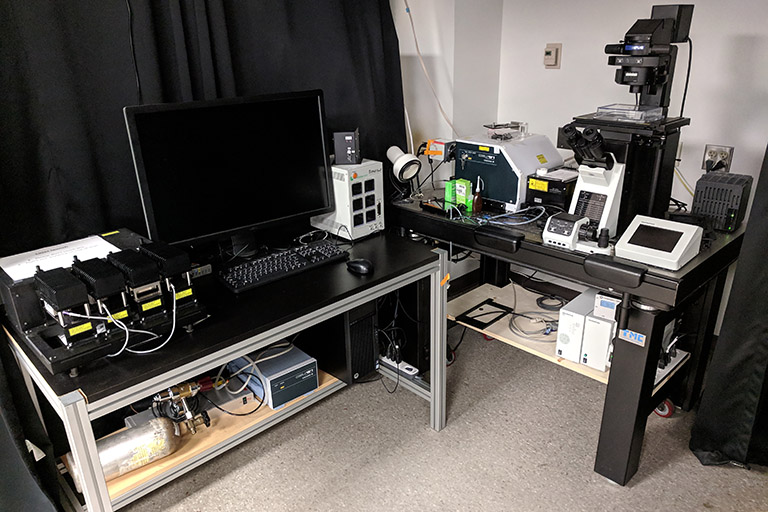
OMX-SR 3D-SIM Super-Resolution System
Offering resolution at twice the diffraction limit of traditional microscopes, the DeltaVision|OMX system is the practical solution for three dimensional super resolution imaging that does not depend on special fluorophores or fluorescent proteins. The OMX is also set-up for TIRF and fast conventional imaging of live cells.
Features of the OMX-SR 3D-SIM Super-Resolution System
- Structured Illumination mode for high-resolution imaging. X,Y resolution to ~ 100nm, Z resolution to ~ 300nm.
- Conventional Illumination mode for fast live cell imaging. Up to 65 frames/second in three colors.
- Lasers lines (405, 488, 561, 642) and and emission filters (419-465, 500-550, 609-654, 665-705) for imaging DAPI, green, red and far red fluorophores.
- Ring TIRF.
- Three PCO Edge 5.5 sCMOS cameras allow simultaneous three color imaging.
- Environmental control for live imaging.
- Controlled by AquireSR software, image processing by SoftWorx imaging software.
More information and resources for the OMX-SR 3D-SIM to IU users
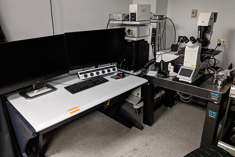
Leica SP8 Scanning Confocal
Installed in January of 2016, the SP8 is a versatile scanning confocal with all the bells and whistles. This system has 4 sensitive HyD detectors and 2 PMT detectors enabling the simultaneous imaging of up to 6 different wavelengths. It has a White Light Laser (WLL) that can provide up to 8 excitation lines between 470 - 670nm) in addition to 405nm and 440nm lasers to provide almost any excitation wavelength and a prism spectrometer to control emission wavelengths to make this a very versatile system compatible with most combinations of fluorophores.
Features of the Leica SP8 Scanning Confocal
- Leica DMi8 inverted microscope platform
- Lasers: WLL (470-670nm), 405nm, 440nm.
- Can simultaneously image fluorescence and brightfield or DIC
- FRAP or FRET fully supported with rapid kinetic analysis
- Picoquant system for FLIM or FCS
- Controlled by Leica LAS-X software
More information and resources for the Leica SP8 microscope to IU users
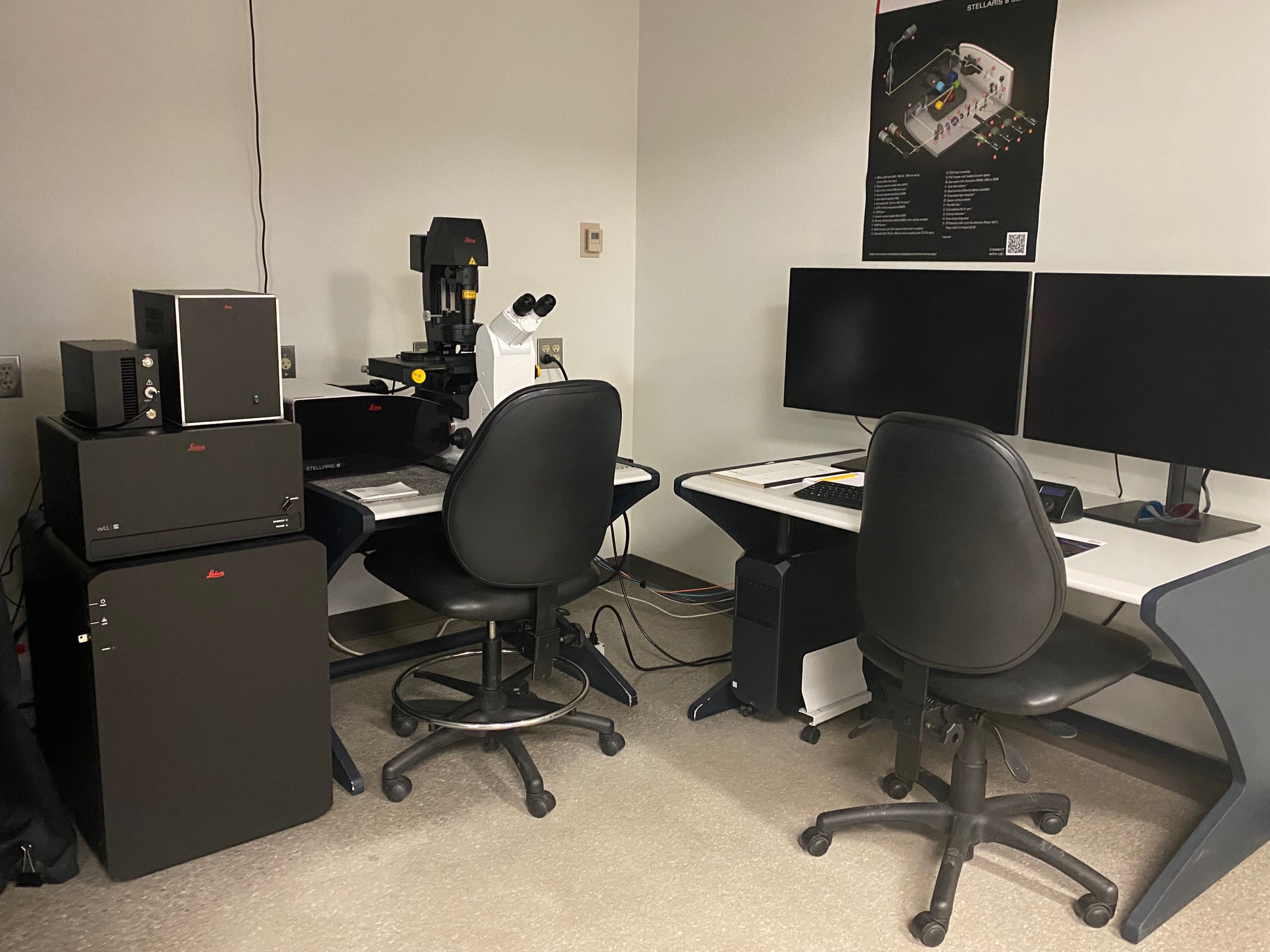
Leica Stellaris 8 FALCON
The new point-scanning confocal system includes an extended white light laser excitation source (select any excitation from 440-790nm), a 405nm excitation laser, an Acusto Optical Beam Splitter (select any fluorescence emission window you want), and state of the art high-sensitivity Power HyD detectors for up to 3 simultaneous fluorescence channels. Special to the Stellaris will be the TauSense toolkit for reducing auto-fluorescence and fully integrated fluorescence lifetime (FLIM) support for quantitative FRET and other applications.
More information and resources for the Leica STELLARIS 8 microscope to IU users
Olympus OSR Spinning Disk Confocal
The OSR microscope achieves super resolution by combining superior optics with the new Yokogawa W1 Spinning Disk to improve signal and reduce noise. In super resolution mode, Airy disk oversampling drives resolution down to 120 nm by capturing subtle high-frequency special components not visible in conventional spinning disk microscopy.
Features of the Olympus OSR Spinning Disk Confocal
- Hammamatsu Fusion BT back illuminated sCMOS camera (100fps full frame, 2304 (H) × 2304 (V), 95%QE)
- 4 laser diodes (445nm, 488nm, 514nm, 561nm)
- Zero-drift autofocus capability
- Can also collect bright-field and DIC images
- Tokai Hit Stage Top Incubation system
More information and resources for the OSR Spinning Disk to IU users
Nikon A1 Scanning Confocal/ TIRF Microscope
Installed May 2012, this versatile system is built on the Nikon Eclipse Ti inverted microscope. It includes the A1 scanning confocal with four lasers that provide five excitation wavelengths, four sensitive fluorescence detectors and can image in both standard or spectral mode. This microscope includes the Perfect Focus system to maintain focus throughout long-term live imaging experiments. Also included are a very nice camera and a TIRF module for imaging high resolution fluorescence in regions very near the coverslip.
Features of the Nikon A1 Scanning Confocal/TIRF Microscope
- Nikon Eclipse Ti inverted microscope platform
- The Nikon Perfect Focus system
- Five laser lines(447, 488, 514, 561, and 640nm) to excite many of the popular fluorophores from CFP to Cy5
- Simultaneous or sequential multichannel imaging
- Standard filter based imaging or Spectral detector for added flexabilty in the separation of fluorophores
- TIRF module with a 100X TIRF objective
- Hammamatsu Orca-Flash 4.0 sCMOS camera
- Controlled by Nikon Elements software
More information and resources for the Nikon A1 microscope to IU users
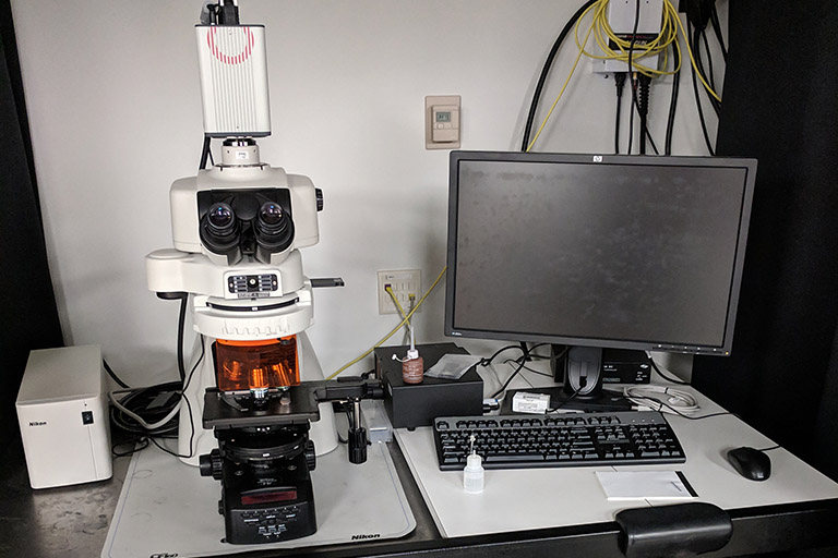
DeltaVision Personal DV
The Applied Precision DeltaVision personalDV Live Cell Imaging System is optimized for deconvolution microscopy. Deconvolution is a software technique that mathematically de-blurs images. This system features a very nice motorized stage for precise control of X, Y, and Z plane movements. Multiple sites on a slide can be marked and revisited.
Features of the DeltaVision Personal DV
- Olympus IX-71 Customized Inverted Microscope Stand
- Photometrics CoolSNAP HQ2 CCD camera
- Insight Solid State Illumination system with filters for DAPI, CFP, GFP/FITC, YFP, RD-TR-PE, mCherry/Alexa594, Cy5 fluorophores
- Can also collect bright-field and DIC images
- WeatherStation Environmental Chamber to control temperature and carbon dioxide levels for live cell imaging
- Controlled by SoftWorx imaging software
More information and resources for the DeltaVision pDV microscope to IU users

Molecular Devices ImageXPress® HCS.ai
The widefield High-Content Screening System captures your imagination with high-quality imaging and analysis, enabling you to acquire the data you need with ease. Empower your team to obtain high-definition images and robust data from a wide range of 2D and 3D cell assays, and leverage the power of AI for deeper insights.
Nikon Eclipse Ni-E
Installed May 2012. The Nikon NiE is a very nice general purpose research microscope. It is equipped for brightfield, DIC, phase and epifluorescence imaging. The microscope has 4X to 100X objectives so a wide range of samples can be accommodated. Epifluorescence imaging on this scope works best with thin, flat specimens.
Features of the Nikon Eclipse Ni-E
- The Nikon NiE is an upright, semi-automated microscope platform
- Hamamatsu Orca-Flash 2.8 sCMOS high resolution camera
- Lumencor SpectraX light source for fluorescence imaging of UV/DAPI, CFP, Green, YFP, Red, and Far-red (CY-5) fluorophores
- Motorized focus control for Z-stack images
- Controlled by Nikon Elements software
More information and resources for the Ni-E system to IU users
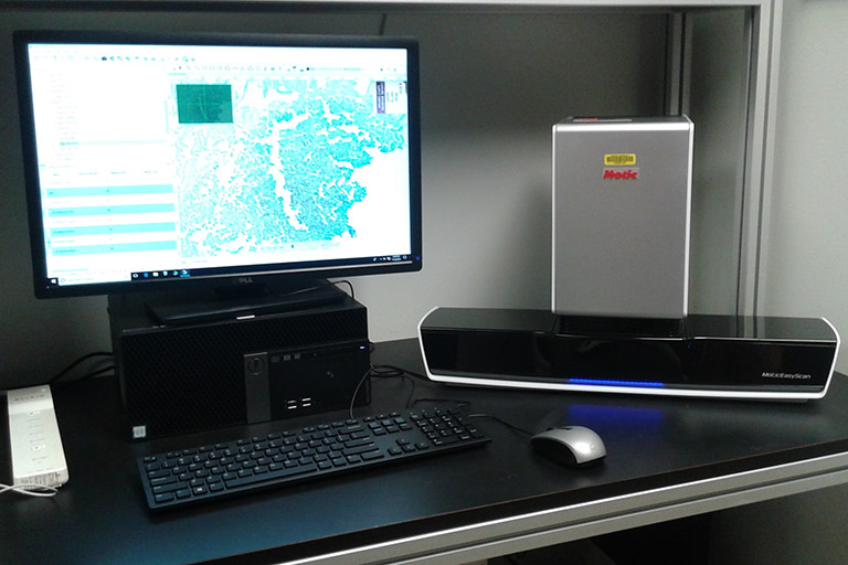
Motic EasyScan
A slide scanning microscope for brightfield imaging. This scope will automatically image whole slides or parts of slides. It is great for histochemically stained samples. Can load up to six slides.
Features of the Motic EasyScan
- 2X - 40X magnification
- Autofocus
- Modes: Normal, High precision, Extended Depth of Field, Z-Stack.
- Slide dimension must be 76mm x 26mm (standard) with a #1.5 coverslip
More information and resources for the Motic EasyScan to IU users
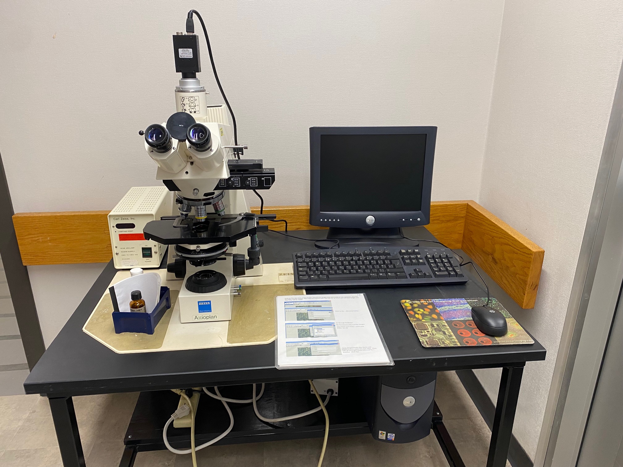
Zeiss Axioplan
The Axioplan is a good general purpose research microscope. It is equipped for brightfield, DIC, and epifluorescence imaging. Images are captured using an IMI Tech IMC-3145FT digital color camera controlled by basic imaging software. This software, while basic, is easy to learn and easy to use. This microscope is on a cart and is available for use in the imaging center or may be used in your lab or classroom.
Features of the Zeiss Axioplan
- The Zeiss Axioplan is an upright microscope platform
- IMI Tech IMC-3145FT color camera
- Hg Lamp illumination with filters for UV/DAPI, Green and Red fluorophores
- Easy to use, a great option for basic imaging or just to look around
- On a cart - may be used in your lab or classroom
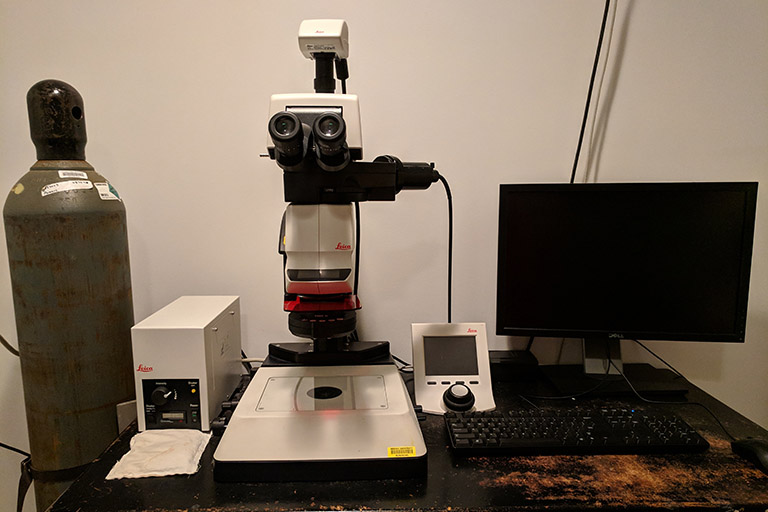
Leica M205FA Stereo Microscope
This is an excellent microscope for a wide range of uses - from specimen preparation and screening to high-resolution imaging of a wide range of samples. It is equipped for brightfield and epifluorescense imaging. Images are captured using the Leica DFC310FX color camera and Leica LAS software.
Features of the Leica M205FA Stereo Microscope
- Hg lamp with filters for UV/DAPI, Green and Red fluorophores
- Magnification from 7.8X to 160X (eyepiece)
- Automated focus and filter controls
Image Processing Workstations
A 64 bit Image Processing workstation with a 30" high resolution screen. Featured software includes:
- Bitplane Imaris 9.9 for 3-D image reconstruction, modeling analysis and presentation. Available licences: MeasurementPro, Coloc, Lineage, Track, Vantage.
- Nikon NIS AR 4.0 Elements for image processing and quantification.
- HuygensEssential 17.1 with Huygens it is possible to perform image deconvolution and restoration, interactive analysis and volume visualization in 2D-4D, multi-channel and time.
- Leica LAS X imaging sofware offline version.
- ImageJ is open source software for processing and analyzing scientific images.
- SymPhoTime 64 2.8 fluorescence liftime imaging and corellation software Powerful 64bit TTTR data acquisition and analysis software. FLIM, fast FLIM, FLIM-FRET. FCS, FCCS, FLCS, PIE-FCS, coincidence correlation, total correlation. FRET, PIE-FRET. Fluorescence time trace analysis, single molecule burst analysis. Anisotropy. TCSPC lifetime fitting with advanced error treatment. User programming script language "STUPSLANG".